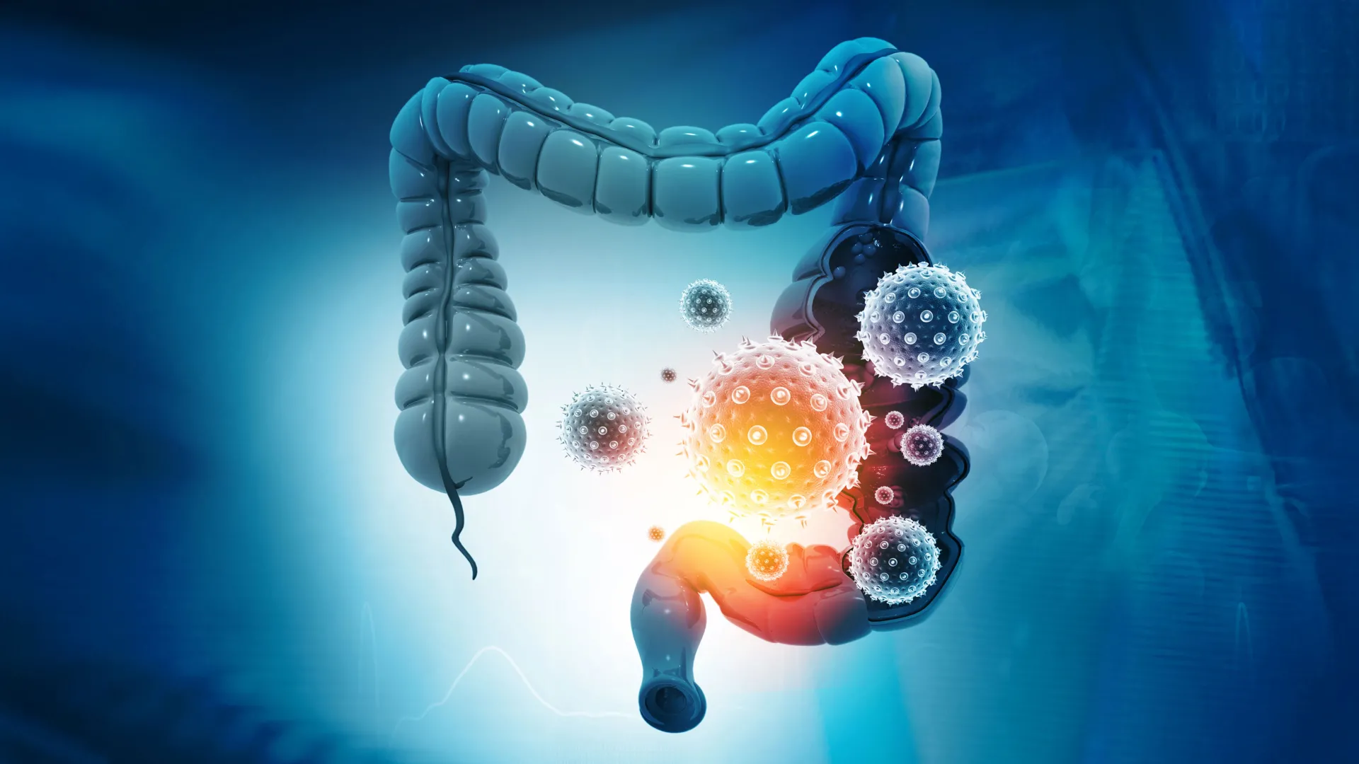
For nearly two decades, the scientific community has grappled with an unsettling paradox regarding human gut health: up to 20 percent of healthy people carry a common bacterium that harbors the potential to destroy the colon’s protective lining and drive tumor formation. While researchers established a definitive link between this bacterium and colorectal cancer years ago, a critical piece of the puzzle remained elusive. Specifically, scientists could not explain how the microbe’s lethal toxin first attached itself to intestinal cells to initiate its destructive assault. Now, an international team of researchers has uncovered that missing step, illuminating a surprising vulnerability that could pave the way for entirely new preventive therapies for colorectal cancer and other gastrointestinal diseases.
The breakthrough, published in the prestigious journal Nature, was spearheaded by a multidisciplinary team at the Johns Hopkins Kimmel Cancer Center, the Bloomberg-Kimmel Institute for Cancer Immunotherapy, and the Johns Hopkins University School of Medicine. By deploying advanced gene-editing technology, the researchers identified a specific host protein that serves as the crucial docking station for the bacterial weapon. Even more promising, the team leveraged this discovery to engineer a molecular decoy that successfully intercepted the toxin in animal models, offering a compelling blueprint for future interventions.
The Chronology of a Decades-Long Scientific Pursuit
To fully grasp the significance of the Johns Hopkins breakthrough, it is necessary to examine the timeline of scientific discovery surrounding toxin-producing gut microbes. The investigation fundamentally centers on Bacteroides fragilis, an anaerobic bacterium native to the human intestinal tract. While non-toxin-producing strains of this bacterium coexist peacefully with their human hosts, contributing to normal digestion, certain strains acquire the genetic capacity to secrete a potent metalloprotease known as Bacteroides fragilis toxin, or BFT.
The journey toward understanding BFT’s role in oncology accelerated dramatically in 2009. In a landmark study published in Nature Medicine, senior author Dr. Cynthia Sears and her colleagues demonstrated that BFT could actively promote the development of colon tumors in animal models. This pivotal work cemented an important epidemiological and mechanistic connection linking chronic intestinal inflammation, microbial metabolites and toxins, and the genesis of colorectal cancer.
Subsequent studies revealed the mechanism by which BFT exerts its cellular damage. Scientists discovered that the toxin targets and cleaves E-cadherin, a vital structural protein responsible for maintaining the adhesion between neighboring cells and preserving the integrity of the intestinal epithelial barrier. When E-cadherin is degraded, the tight junctions of the colon fail, creating a compromised barrier that permits luminal bacteria and inflammatory triggers to infiltrate deeper tissues, thereby fueling chronic inflammation and tumorigenesis.
Despite these revelations, a foundational question persisted: how did the toxin physically engage the cellular machinery? Although BFT efficiently cleaved E-cadherin, experimental observations showed that the toxin did not bind directly to E-cadherin itself. Researchers long suspected the existence of an intermediary receptor molecule that guided the toxin to its destination, but repeated attempts over nearly twenty years to identify this elusive molecule ended in frustration.
Unlocking the Secret With CRISPR Technology
The turning point arrived when the Johns Hopkins team partnered with researchers at Harvard Medical School to deploy CRISPR-Cas9, a revolutionary gene-editing tool capable of systematically inactivating specific genes to observe the functional consequences. Maxwell White, an M.D./Ph.D. candidate in Dr. Sears’ laboratory, spearheaded a genome-wide CRISPR screening effort in close collaboration with the laboratory of Dr. Matthew Waldor at Harvard.
The researchers systematically disabled individual genes within colon epithelial cells—the specialized cells forming the inner lining of the human colon—to identify which genetic components were strictly required for BFT to attach and inflict cellular harm. Through high-throughput screening, one gene emerged as a resounding and unambiguous top hit. This gene encoded claudin-4, a transmembrane protein instrumental in the formation of tight junctions that seal the spaces between adjacent epithelial cells.
When the team experimentally silenced the claudin-4 gene, the dynamic shifted entirely: BFT could no longer bind to the surface of the colon cells. Deprived of its initial docking site, the toxin was rendered incapable of reaching or cleaving its downstream target, E-cadherin. This definitive experiment proved that claudin-4 functions as the long-sought primary receptor for the bacterial toxin, acting as the mandatory gateway for the pathogen’s attack.
"It took a while to get the assay working and validate the approach, but once we were able to do the screen, claudin-4 was a clear, resounding top hit," remarked Maxwell White regarding the breakthrough. "That was an exciting moment."
An Unexpected Paradigm Shift in Bacterial Pathogenesis
The identification of claudin-4 as the BFT receptor stunned the scientific community. Prior to this discovery, Dr. Sears and other gastrointestinal researchers had operated under the working hypothesis that BFT would naturally interact with a cell signaling receptor, such as a G protein-coupled receptor (GPCR). GPCRs act as primary communication switches on cell surfaces, allowing cells to perceive and adapt to extracellular environmental cues.
Claudin-4, conversely, serves a vastly different biological role. Rather than functioning as a signaling hub, its primary responsibility is structural: anchoring adjacent cells together to maintain tissue architecture and regulate paracellular permeability. Furthermore, the discovery underscores an unusual operational model for a bacterial protease. While most proteases directly engage and cleave their functional targets, BFT relies on a structurally distinct receptor to anchor itself to the cell membrane before it can execute its enzymatic destruction of E-cadherin. Comprehensive literature reviews conducted by the research team confirmed that no other known bacterial toxin utilizes this exact mechanistic strategy.
To cement these genetic findings with robust structural evidence, the Johns Hopkins researchers teamed up with structural biologists F. Xavier Gomis-Rüth and Ulrich Eckhard at the Molecular Biology Institute of Barcelona. Utilizing advanced biophysical assays designed to measure molecular interactions, the team proved that BFT and claudin-4 form a tightly bound macromolecular complex in vitro, with each individual toxin molecule pairing precisely with one receptor molecule.
Engineering a Molecular Decoy to Thwart Infection
Equipped with the knowledge of how BFT docks to human intestinal cells, the research team quickly pivoted from basic discovery to translational application. If BFT requires claudin-4 to initiate its cascade of cellular destruction, the scientists reasoned that introducing a decoy molecule could successfully lure the toxin away from vulnerable host tissues.
To test this hypothesis, the team engineered a soluble molecular decoy protein incorporating the specific binding sequences of claudin-4. This decoy was designed to act as a molecular sponge, capturing circulating BFT in the intestinal lumen before the toxin could make contact with endogenous cellular receptors on the colon’s epithelial lining.
Collaborating with the laboratory of Dr. Min Dong at Harvard Medical School—including researcher Kang Wang—the Johns Hopkins team tested this therapeutic strategy in vivo using mouse models. The results exceeded expectations: rather than binding to the claudin-4 receptors embedded in the colon wall, the circulating BFT preferentially attached to the soluble decoy proteins. By intercepting the pathogen’s initial attachment phase, the decoy successfully protected the mice from the destructive inflammatory and barrier-disrupting effects of the toxin.
"This approach could be iterated upon with small molecules or other biologics that have better pharmacological properties," noted White, outlining the future trajectory of the research. The Johns Hopkins laboratory is currently evaluating various small-molecule compounds and biologic candidates to optimize the stability, pharmacokinetic profile, and delivery mechanisms of the decoy strategy for potential human clinical translation.
Broader Implications for Diagnostics and Cancer Prevention
While the development of a molecular decoy represents a monumental leap forward in preclinical gastroenterology, researchers emphasize that significant milestones remain before human clinical trials can begin. Although the mouse studies definitively demonstrated protection against acute tissue damage and barrier degradation, the experiments did not explicitly prove that the decoy strategy prevents the long-term development of colorectal cancer. Additionally, the researchers have yet to capture the exact three-dimensional atomic structure of the BFT-claudin-4 complex—a feat that even cutting-edge artificial intelligence platforms, including AlphaFold, have not yet fully resolved.
Despite these hurdles, the discovery resolves a major biomedical mystery that has persisted for nearly two decades. By illuminating the precise molecular handshake that initiates bacterial pathogenesis in the lower gastrointestinal tract, the team has opened new vistas for preventive oncology and infectious disease management.
Reflecting on the broader impact of the study, senior author Dr. Cynthia Sears highlighted the wide-ranging potential of the findings. "We’ve made several attempts over time to identify the receptor, so this is an exciting moment," said Dr. Sears, who holds the title of Bloomberg-Kimmel Professor of Cancer Immunotherapy and professor of medicine at Johns Hopkins. "Understanding how bacterial toxins work can open doors to new approaches for detection and therapy for associated diseases, including diarrhea, colorectal cancer and bloodstream infections."
As the scientific community moves forward, this foundational discovery provides a clear roadmap for intercepting microbial pathogens at the earliest possible stage. By neutralizing threats before they breach the cellular gates, future therapies may effectively neutralize the carcinogenic potential of common gut bacteria, offering robust new layers of defense for human gastrointestinal health.
Additional researchers contributing to this landmark study include Jason Chen, Shaoguang Wu, Abby L. Geis, and Jessica Queen of Johns Hopkins University, alongside Hailong Zhang, Karthik Hullahalli, and Jie Zhang of Harvard Medical School. This investigation was supported by funding from the Bloomberg-Kimmel Institute for Cancer Immunotherapy, Janssen Research and Development, Cancer Research UK, the Howard Hughes Medical Institute, and the National Institutes of Health under grant numbers R01 AI042347, R01 NS080833, R01 NS117626, R01 AI170835, and R01 AI189789.