For more than seventy years, the pharmaceutical compound 6-thioguanine (6-TG) has stood as a cornerstone in the therapeutic arsenal against leukemia. As a member of the thiopurine class of medications, its primary clinical application has been to disrupt the replication of rapidly dividing malignant cells, offering a vital lifeline to generations of cancer patients. Yet, despite its extensive documentation and widespread clinical utility, the precise molecular mechanisms governing patient response have remained stubbornly opaque. Oncologists and pharmacologists have long grappled with a fundamental biological paradox: while some cellular populations succumb rapidly to the cytotoxic assault of 6-TG, others exhibit robust resilience, managing to withstand the treatment entirely.
An international research consortium, spearheaded by the CeMM Research Center for Molecular Medicine of the Austrian Academy of Sciences in collaboration with the University of Oxford, the Weizmann Institute of Science, and the University of Dundee, has now upended conventional pharmacological assumptions. In a breakthrough study that bridges structural biology and targeted therapeutics, the scientific team has identified an unexpected cellular protagonist dictating this drug response: a protein known as NUDT5. Rather than operating through its traditional chemical pathways, NUDT5 appears to influence cellular survival through a physical, structural role that challenges traditional drug-design paradigms and opens new avenues for optimizing cancer chemotherapy.
A Paradigm Shift in Protein Function: Beyond Catalytic Activity
The discovery did not emerge in a vacuum; it builds directly upon foundational insights published recently in the journal Science by the Kubicek and Huber laboratories. That prior research established a paradigm-shifting concept regarding NUDT5: the protein possesses a critical intracellular role entirely independent of its conventional enzymatic activity. While biochemistry textbooks traditionally define proteins like NUDT5 as catalysts designed to drive specific chemical reactions by converting substrates into products, this recent work revealed a vastly different dimension. NUDT5 can also function as a molecular scaffold—a structural anchor that physically organizes and coordinates cellular metabolism.
It is this non-enzymatic, structural function that appears to dictate how cells respond to 6-thioguanine. For the researchers at CeMM, the revelation came as a surprise. "We initially expected that NUDT5 would influence 6-TG through its enzymatic activity," explains Tuan-Anh Nguyen, co-first author of the study from CeMM. "Instead, we found that inhibiting the enzyme had little effect. What mattered was whether the protein itself was present."
This distinction carries profound implications for drug development. For decades, the pharmaceutical industry has relied predominantly on small-molecule inhibitors designed to fit snugly into the active sites of target enzymes, blocking their catalytic functions. However, when the researchers applied conventional inhibitors to block NUDT5’s catalytic capabilities, cellular sensitivity to 6-TG remained largely unaltered. The enzyme’s chemical work was not what mattered; its physical existence within the cell architecture was the decisive variable.
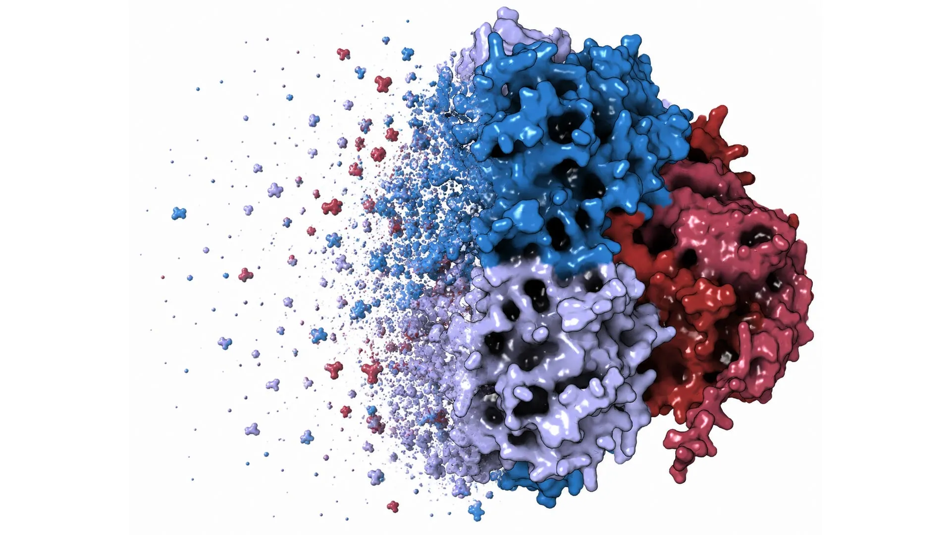
Deploying Targeted Protein Degradation: The Birth of dNUDT5
To definitively prove that the physical presence—rather than the catalytic function—of NUDT5 drove the cellular response to 6-TG, the research team had to move beyond inhibition. They turned to an cutting-edge methodology that has rapidly gained traction in modern chemical biology: targeted protein degradation.
Unlike traditional inhibitors that merely silence a protein’s catalytic engine while leaving the physical structure intact inside the cell, targeted protein degradation co-opts the cell’s own waste-disposal machinery to completely eliminate the target protein. To achieve this, a medicinal chemistry program led by the Huber laboratory at the University of Oxford embarked on a rigorous design campaign.
"We developed a cell-based platform to accelerate the discovery of NUDT5 degraders," explains Anne-Sophie Marques, a first author whose contributions at Oxford were instrumental to the project. This innovative platform guided medicinal chemistry efforts to synthesize a diverse library of highly selective NUDT5 degraders, culminating in the creation of dNUDT5, the most potent and active degrader in the series. Simultaneously, the team engineered matched control compounds capable of binding to NUDT5 without triggering its destruction, ensuring rigorous experimental validation.
When the researchers systematically compared the effects of these newly minted degraders against conventional enzymatic inhibitors, the biological divergence was stark. While conventional inhibitors failed to shift the needle on 6-TG toxicity, complete removal of the NUDT5 protein via dNUDT5 conferred a striking protective effect, shielding the cells from the drug’s lethal cytotoxicity. Subsequent genetic knockout experiments yielded identical conclusions, cementing the validity of the phenomenon.
"Chemical degraders give us a way to separate what a protein does as an enzyme from what it does as a physical presence in the cell," notes Professor Kilian Huber of the Centre for Medicines Discovery at the University of Oxford and co-corresponding author of the study. "In this case, that distinction was decisive: removing NUDT5 revealed biology that conventional inhibitors missed."
Unraveling the Thiopurine Network: The Counterbalance of NUDT5 and NUDT15
As the empirical data accumulated, the research team observed a dose-dependent protective effect. Ludwig Bauer, another first author of the study, recalls the moment the breakthrough became apparent: "As the results came in, it became immediately clear that the dNUDT5 was protecting cells from 6-thioguanine toxicity in a dose-dependent manner. That was an incredibly exciting moment."
Intriguingly, the investigation also uncovered a complex biological crosstalk between NUDT5 and another protein well-known to clinical pharmacologists: NUDT15. For years, medical science has recognized that genetic variations in NUDT15 significantly influence how individual patients metabolize and respond to thiopurine drugs, often dictating whether a standard dose will lead to severe toxicity or therapeutic failure.
Yet, despite belonging to the same protein family, NUDT5 and NUDT15 appear to exert opposing forces on cellular sensitivity to 6-TG. While the loss or reduction of NUDT15 typically sensitizes cells, making them more vulnerable to 6-thioguanine, the reduction or removal of NUDT5 drives the opposite phenotypic response, rendering cells markedly more resistant. These opposing trajectories indicate that thiopurine response is not governed by a linear pathway, but rather by a sophisticated network of protein-protein interactions and structural balances that can push cellular fate in divergent directions.
"Our results show that proteins can have important biological functions that are completely independent of their enzymatic activity," summarizes Stefan Kubicek, Principal Investigator at CeMM and corresponding author of the study. "By removing NUDT5 rather than simply inhibiting it, we were able to uncover a hidden layer of biology that helps determine how cells respond to a clinically important drug."
Broader Impacts and Future Implications for Cancer Therapeutics
While the researchers are quick to emphasize that these fundamental discoveries do not immediately translate into a novel clinical treatment at the bedside, the implications for translational oncology and drug discovery are profound.
First, the study provides a compelling explanation for the long-standing variability observed in thiopurine treatment outcomes. By illuminating the non-catalytic structural role of NUDT5, scientists now have a new theoretical framework to investigate why certain leukemia cells evade the cytotoxic mechanisms of 6-TG. This could eventually inform the development of predictive biomarkers or combination therapies designed to bypass cellular resistance mechanisms.
Second, the research serves as a powerful validation of targeted protein degradation as an indispensable tool in modern drug discovery. By demonstrating that traditional enzymatic assays and inhibitors can leave vital pharmacological blind spots, the study underscores the necessity of exploring non-catalytic protein functions. Proteolysis-targeting chimeras (PROTACs) and related degrader technologies allow scientists to probe biological spaces that were previously deemed undruggable or invisible to classical pharmacology.
The collaborative effort behind these findings was made possible through robust international funding and institutional support, reflecting the high priority assigned to cutting-edge molecular research in Europe. Financial backing was provided by the European Research Council (ERC) under the European Union’s Horizon 2020 research and innovation program, the Austrian Science Fund (FWF), the Vienna Science and Technology Fund (WWTF), and the Marie Skłodowska-Curie Actions Postdoctoral Fellowships program. Additional support came from the Innovative Medicines Initiative 2 Joint Undertaking (IMI2 JU), the Wellcome Trust, Merck Sharp & Dohme Corp., and Janssen Pharmaceutica NV.
As the scientific community continues to digest these findings, the study stands as a testament to the power of questioning long-held dogmas. By looking past an enzyme’s chemical job description and focusing instead on its physical architecture within the cell, researchers have unlocked a hidden layer of cellular biology—offering a renewed lens through which to view one of medicine’s oldest cancer-fighting drugs.