In a monumental breakthrough for musculoskeletal medicine, a collaborative team of researchers at Weill Cornell Medicine and the Hospital for Special Surgery (HSS) has successfully identified a previously elusive population of stem cells responsible for generating the body’s tendons and ligaments. This foundational discovery not only fills a decades-long knowledge gap in developmental biology but also sheds light on the root causes of lumbar spinal stenosis, a debilitating condition affecting more than 100 million people worldwide. Published in the peer-reviewed scientific journal Cell, the findings introduce a new cellular paradigm that could pivot spinal care away from invasive surgeries and toward targeted pharmacological interventions.
For years, the cellular origins of tendons and ligaments—the fibrous connective tissues that anchor muscles to bones and stabilize joints—remained shrouded in mystery. Unlike bone tissue, which has clear structural markers and well-documented progenitor cells, tendons and ligaments are densely populated with visually similar fibroblast-like cells. This cellular homogeneity made it exceptionally difficult for scientists to isolate a single, pure population capable of true stemness: the dual capacity to self-renew indefinitely while differentiating into the full spectrum of specialized mature tissue cells.
The Research Journey and Chronological Discovery
The identification of this universal tendon and ligament stem cell was the culmination of years of meticulous investigation by the research team led by Dr. Matthew Greenblatt, associate professor of pathology and laboratory medicine at Weill Cornell Medicine and a pathologist at NewYork-Presbyterian/Weill Cornell Medical Center, alongside Dr. Sravisht Iyer, an associate professor of orthopedics at Weill Cornell Medicine and a spine surgeon at HSS.
The timeline of this discovery builds upon Dr. Greenblatt’s extensive prior work in skeletal stem cell biology. In 2018, Dr. Greenblatt’s laboratory made headlines by identifying the specific skeletal stem cell responsible for initiating fracture repair within the periosteum, the outer layer of bone. Subsequent studies by the same group uncovered progenitor cells involved in the embryonic and postnatal formation of the skull and spine. However, isolating an equivalent population for soft connective tissues proved to be a formidable scientific hurdle.
To overcome the challenge of cellular heterogeneity, the research team deployed advanced single-cell RNA sequencing technologies. By analyzing thousands of individual cells harvested from murine models, the scientists meticulously sorted them by genetic expression profiles, isolating the precise population that exhibited hallmark traits of stemness. Subsequent lineage-tracing experiments confirmed that these rare cells reside within specialized anatomical niches inside tendons and ligaments, functioning as an internal reservoir dedicated to tissue maintenance and repair.
Translating Animal Models to Human Pathology
Following the successful identification of the stem cells in mice, the research team transitioned to human tissue samples to determine if the findings were clinically relevant. Under strict institutional protocols and with informed consent, human ligament tissues were harvested during routine spinal decompression surgeries performed by Dr. Iyer.
Led by first author Dr. Lingling Hu, a postdoctoral fellow operating jointly in the Greenblatt and Iyer laboratories, the team isolated corresponding human cells and verified their dual functionality: the capacity for self-renewal and the generation of functional ligament lineage cells. Furthermore, through expansive mapping across various anatomical sites—including the patellar ligament of the kneecap and the Achilles tendon—the researchers confirmed that these progenitor cells are not localized solely to the spinal column.
"We looked in the kneecap ligament; we looked at the Achilles tendon; and everywhere we looked, we found this cell," Dr. Greenblatt noted. "So, we think this is the universal stem cell for tendons and ligaments throughout the body."
Implications for Lumbar Spinal Stenosis
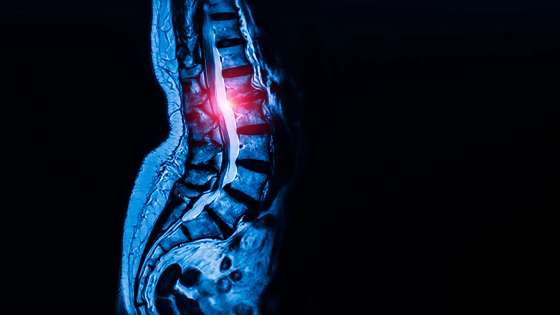
With the foundational biology established, the researchers shifted their focus to pathological conditions, specifically lumbar spinal stenosis. Affecting an estimated 103 million individuals globally, spinal stenosis is a degenerative condition characterized by the abnormal thickening of spinal ligaments and bone structures. This tissue hypertrophy progressively narrows the spinal canal, placing immense physical compression on central nerve roots. Patients frequently experience chronic lower back pain, radiating numbness, tingling, and severe mobility impairments that drastically diminish their quality of life.
Historically, therapeutic options for severe spinal stenosis have been limited. Once conservative treatments such as physical therapy and corticosteroid injections fail, patients are routinely funneled toward invasive surgical procedures, such as laminectomies or spinal fusions, to manually relieve nerve compression.
Seeking a more proactive, mechanistically driven approach, the research team compared stem cell populations extracted from spinal ligaments of patients undergoing surgery for spinal stenosis against control samples harvested from patients with herniated discs who exhibited no signs of stenosis.
The comparative analysis revealed a striking biological disparity: ligaments derived from spinal stenosis patients contained a significantly higher concentration of the newly identified stem cells. When these pathological stem cells were transplanted into murine models, they drove accelerated and excessive production of tendon and ligament cells compared to control cells.
Decoding the Cellular Mechanism: Calcium Signaling
Determined to understand the biochemical drivers behind this overactivity, the investigators examined intracellular communication pathways within the stem cells. The analysis revealed that stem cells sourced from spinal stenosis patients exhibited hyperactive calcium signaling—a fundamental cellular messaging system that governs growth, gene expression, and tissue proliferation.
To test causality, the research team utilized genetic techniques to artificially increase calcium signaling within healthy stem cells. This manipulation successfully induced excessive tissue growth mirroring the pathology of spinal stenosis. Conversely, when the researchers pharmacologically or genetically suppressed calcium signaling in a mouse model of the disease, the abnormal, excessive tissue proliferation was effectively halted.
This pivotal mechanistic insight suggests that hyperactive calcium signaling acts as the primary molecular engine driving the overgrowth of ligaments in spinal stenosis. Consequently, the pathway opens an entirely novel avenue for non-surgical therapeutic intervention.
A Potential Repurposing of Existing Medications
Because calcium signaling plays a central role in vascular biology and cardiac function, a well-established class of therapeutics already exists: calcium channel blockers. These medications are widely prescribed globally to manage systemic hypertension and various cardiovascular disorders.
The discovery that calcium channel blockers could potentially regulate the hyperactive stem cells responsible for ligament thickening raises the tantalizing prospect of drug repurposing. If clinical trials confirm safety and efficacy, physicians may one day be able to treat the root cellular mechanisms of spinal stenosis using oral medications, potentially delaying or entirely averting the need for major corrective surgery.
"This is probably the first work that’s shown a potential therapeutic target for one of the most common spine conditions in the world," Dr. Iyer emphasized, highlighting the transformative potential for patient care pathways. "Identifying these specialized stem cells unlocks a new area of research that allows us to address this disease much more mechanistically, rather than just waiting until a patient’s condition worsens and requires surgery to relieve the nerve compression."
Broader Medical Horizons and Future Research Directions
While the immediate clinical applications focus heavily on lumbar spinal stenosis, the discovery of a universal tendon and ligament stem cell carries sweeping implications across multiple disciplines of orthopedic medicine, regenerative medicine, and genetic pathology.
Because these progenitor cells appear to be ubiquitous throughout the musculoskeletal system, defects or dysregulations within this specific stem cell population are likely implicated in a wide array of chronic connective tissue disorders and traumatic injuries. Future research initiatives are expected to explore the involvement of these cells in notoriously difficult-to-heal soft tissue injuries, including chronic rotator cuff tears, severe Achilles tendon ruptures, and complications arising from ligament reconstruction surgeries.
Furthermore, the research team plans to investigate whether aberrant activity within these same stem cell reservoirs contributes to systemic genetic conditions such as Marfan syndrome, a hereditary disorder that severely compromises connective tissue integrity throughout the entire body.
As the medical community digests the implications of this study, validation through rigorous human clinical trials will be the necessary next step to translate these bench-science discoveries into viable patient treatments. Nonetheless, by illuminating the cellular architects behind tendons, ligaments, and spinal pathology, this collaborative effort between Weill Cornell Medicine and the Hospital for Special Surgery marks a defining milestone in modern musculoskeletal research.
Funding and Institutional Support
This extensive research endeavor was made possible through the generous financial backing of numerous institutional and philanthropic organizations. Major support was provided by the Marfan Foundation’s Victor A. McKusick Fellowship, a Kellen Scholars Award, the Children’s Tumor Foundation, the Arthritis National Research Foundation, a Jumpstart award from Weill Cornell Medicine, and the National Research Foundation of Korea (NRF) funded by the Ministry of Education. Additional funding sources included the National Institutes of Health (grants T32-AR078751 and T32-AR071302-07), a MIND Prize from the Pershing Square Foundation, a Mary Kay Ash Foundation Award, an Innovator Award from the Marfan Foundation, and a Burroughs Wellcome Fund Career Award for Medical Scientists.