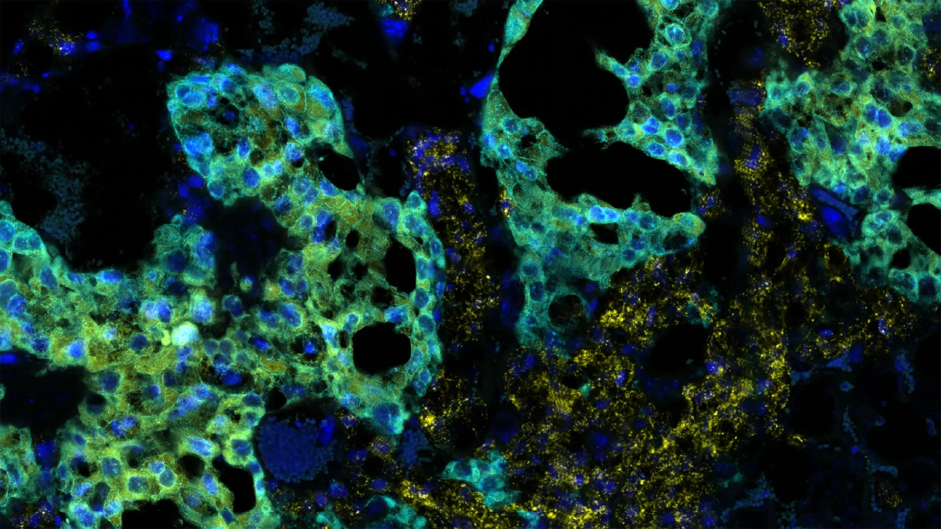
The quest to fully understand the origins of autism spectrum disorder (ASD) has historically focused heavily on neurological development, genetic architecture, and postnatal behavioral observations. However, a compelling body of emerging research suggests that the earliest roots of developmental divergence may be planted long before birth, extending deep into the gestational environment. In a major scientific development, researchers at the Cold Spring Harbor Laboratory (CSHL) have published findings that establish a surprising, highly specific causal chain linking maternal immune activation, structural damage to the placenta, and the subsequent emergence of autism-like traits—exclusively in male subjects.
The study, led by CSHL Associate Professor Lucas Cheadle alongside postdoctoral researcher Irene Sanchez Martin, pivots away from traditional brain-centric models of autism research. By investigating the intricate physiological dialogue between a mother and her developing fetus, the CSHL team has uncovered a critical window of vulnerability during gestation. These insights not only challenge long-held paradigms regarding when and how developmental disruptions begin, but they also offer a fresh perspective on one of the most persistent epidemiological puzzles in modern medicine: why autism is diagnosed significantly more often in males than in females.
While the experiments were conducted using murine models and do not yet definitively prove identical pathways in humans, the implications of the research stretch far across the landscapes of prenatal care, immunology, and developmental neurobiology. As scientists worldwide race to map the multifaceted etiologies of ASD, this study serves as a vital reminder that the gateway to neurological development may be guarded by the placenta.
Unraveling the Gestational Timeline and Windows of Vulnerability
For decades, developmental biologists and immunologists have studied maternal immune activation (MIA)—a phenomenon where an expectant mother’s immune system mounts a defense against a pathogen, such as a virus or bacteria. While the mother’s body successfully clears the infection, the inflammatory cytokines released during this immune response can occasionally cross the placental barrier, inadvertently altering the trajectory of fetal development.
To dissect the precise mechanics of this process, Cheadle and his colleagues designed a series of controlled experiments. They administered a specialized synthetic substance to pregnant mice designed to precisely mimic the immune system’s physiological reaction to a viral infection, completely avoiding the confounding variables of an active, replicating virus. Crucially, the researchers tested this immune challenge at multiple, distinct stages of the murine gestation cycle to determine if susceptibility varied over time.
The results revealed a remarkably narrow and consequential window of vulnerability. When maternal immune activation was triggered precisely 12.5 days into the mouse pregnancy—roughly the midpoint of gestation—the consequences were immediate and profound. According to Irene Sanchez Martin, approximately one-third of the developing fetuses exhibited clear, measurable developmental abnormalities within a staggering 24-hour window following the simulated infection.
This timeline is particularly noteworthy because it captures developmental disruption at an earlier stage than researchers had ever previously documented in MIA models. By pushing the observational timeline back to these critical early embryonic hours, the CSHL team demonstrated that the biological cascades leading to neurodevelopmental variations are initiated much earlier in gestation than traditional diagnostic models have assumed.
The Placental Gateway: Spongiotrophoblasts Under Siege
To understand why only a fraction of the fetuses suffered abnormalities while others appeared resilient, the CSHL researchers turned their analytical focus toward the placenta. Long viewed merely as a passive organ for nutrient exchange and waste removal, contemporary immunology recognizes the placenta as a sophisticated, dynamic neuro-immune interface that actively regulates the fetal environment.
Within the mouse placenta, the researchers zeroed in on a specialized population of cells known as spongiotrophoblasts. These cells occupy a critical anatomical zone where maternal blood vessels interface directly with fetal tissues, creating a functional bridge between two distinct biological systems.
Upon examining the placental tissue of the affected fetuses, the researchers discovered visible, extensive cellular damage localized specifically within the spongiotrophoblast layer. Conversely, in fetuses that continued to develop normally, this specialized cellular zone remained healthy and intact.
The functional implications of this discovery are profound. Spongiotrophoblasts play an irreplaceable role in immunological tolerance, acting as a peacekeeping force that prevents the mother’s immune system from recognizing the fetus—which carries foreign paternal genetic material—as an invader that must be rejected. When maternal immune activation disrupts or damages these specialized cells, that vital protective barrier is compromised. The maternal inflammatory signals can then penetrate deeper, directly impacting the developing fetal systems in a destructive manner.
This finding reframes the placenta not merely as a casualty of systemic inflammation, but as an active participant—and a potential gatekeeper—in the pathogenesis of neurodevelopmental conditions like autism.
The Sex-Specific Divide: Decoding the Male Vulnerability
Perhaps the most striking and clinically relevant discovery of the CSHL study was the stark dichotomy observed between male and female fetuses. Across every experimental iteration, autism-like behavioral and physiological traits manifested exclusively in the male subjects. The female fetuses, subjected to the exact same maternal immune activation and gestational timelines, continued to develop normally without showing analogous disruptions.
This pronounced sex disparity mirrors human epidemiological data, where autism spectrum disorder is diagnosed in males at a rate roughly four times higher than in females. For years, researchers have debated the origins of this skew, proposing hypotheses ranging from diagnostic bias to sex-specific genetic buffering. However, the CSHL findings introduce a radical new timeline to the discussion.
The sex-based differences in vulnerability emerged at a developmental stage that precedes hormonal surges and long before the onset of sexual differentiation in the brain and other key anatomical regions.
"It’s really fascinating, because we’re looking at an age which precedes hormonal surges," Cheadle noted, highlighting the unexpected nature of the data. "It precedes sexual differentiation in the brain and many other regions. So, it’s surprising there would be a sex-based difference this early."
This temporal paradox suggests that the mechanisms driving male vulnerability are rooted in fundamental, foundational cellular and genetic differences that exist prior to endocrine-driven sexual maturation.
Investigating the Female Advantage
The immunity of the female fetuses to the inflammatory challenge has opened up an entirely new avenue of investigation within Cheadle’s laboratory. Scientists are now aggressively pursuing two primary hypotheses to explain why female embryos appear naturally fortified against maternal immune activation.
The first hypothesis centers on embryonic proteins. It is possible that male embryos express specific proteins or surface markers that inadvertently provoke a more aggressive, damaging secondary reaction from the maternal immune system following a viral challenge.
The second hypothesis posits that female embryos possess an intrinsic, genetically encoded protective mechanism—perhaps linked to X-chromosome gene dosage or unique cellular stress-response pathways—that allows them to withstand high levels of gestational inflammation during sensitive developmental epochs.
"A protective mechanism may exist within the female that could be harnessed to help the male," Cheadle explained, outlining the translational potential of ongoing research. "We’re trying to understand how autism emerges in both sexes. The evidence says that there are potentially different mechanisms."
By identifying the exact molecular pathways that grant female fetuses this apparent resilience, researchers hope to discover novel therapeutic targets. If scientists can decode the female protective mechanism, it may eventually be possible to engineer pharmacological or biological interventions that mimic this protection in vulnerable male pregnancies.
Broader Implications and Future Directions in Autism Research
As the scientific community digests the CSHL findings, the broader implications for prenatal health and neurodevelopmental research are substantial. While the study was conducted entirely in murine models—a necessary step for isolating variables and examining tissue at precise gestational moments—the overarching conceptual framework demands a re-evaluation of how human pregnancies are monitored and managed during viral outbreaks or systemic inflammatory events.
Historically, clinical focus during pregnancy has prioritized preventing direct pathogen transmission to the fetus, such as in the cases of Zika or rubella. However, studies like this underscore the reality that maternal systemic inflammation alone—even in the absence of a persisting viral infection within the fetus—can alter fetal neurodevelopment via placental mediation.
Looking forward, Cheadle’s research team plans to deepen their comparative analysis of male and female developmental pathways. Their ultimate objective is to pinpoint the exact molecular signals exchanged between the damaged placenta and the fetal brain, mapping out the complete chain of events from maternal immune response to observable behavioral traits.
Such insights could eventually revolutionize the field of early intervention. By recognizing the placenta as an early warning system and a primary locus of vulnerability, medical researchers may one day develop diagnostic biomarkers capable of assessing placental health and inflammatory damage in real-time during pregnancy.
While much work remains to translate these animal models into clinical applications for humans, the CSHL study marks a paradigm shift. By looking beyond the confines of the brain and directing their gaze toward the placenta, researchers have unlocked a promising new chapter in the ongoing effort to understand, diagnose, and eventually support individuals on the autism spectrum.