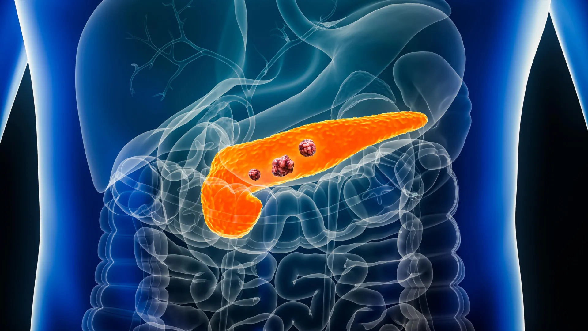
Pancreatic ductal adenocarcinoma remains one of the most lethal and elusive malignancies in modern oncology, frequently claiming lives because it is typically identified only after reaching an advanced, untreatable stage. However, a breakthrough artificial intelligence model known as REDMOD has demonstrated the capacity to spot imperceptible, microscopic alterations in pancreatic tissue long before conventional imaging technologies or experienced clinicians can detect them. Published recently in the peer-reviewed medical journal Gut, this development introduces a paradigm shift in how medicine might approach one of the deadliest forms of cancer by moving the timeline of detection from reactive symptom management to proactive, preclinical intervention.
The Core Innovation: How REDMOD Works
The primary obstacle in treating pancreatic ductal adenocarcinoma lies in the anatomy and behavior of the pancreas itself. Situated deep within the abdomen, the organ is surrounded by complex vascular structures and neighboring organs, making early visualization notoriously difficult. Furthermore, early-stage tumors typically generate minimal symptoms, allowing the malignancy to progress rapidly and silently. Standard computed tomography (CT) scans often fail to capture the earliest structural anomalies because the tissue modifications are too fine for the human eye to isolate amidst normal biological noise.
To overcome these diagnostic barriers, an interdisciplinary team of researchers developed the Radiomics-based Early Detection MODel, or REDMOD. Unlike conventional AI systems that simply scan for visible masses, REDMOD utilizes advanced radiomics—the mathematical extraction of quantitative data from medical images. The algorithm meticulously analyzes microscopic patterns in tissue texture that remain entirely invisible on standard CT scans.
Additionally, REDMOD automates a critical preliminary step by identifying, outlining, and segmenting the pancreas from adjacent structures. In traditional clinical settings, radiologists must manually trace the boundaries of the organ, a labor-intensive process that introduces human error and inter-observer variability. By standardizing this segmentation process, REDMOD ensures consistent evaluation across diverse imaging datasets, eliminating subjective discrepancies that often hinder early diagnosis.
A Retrospective Chronology: Tracing the AI’s Lead Time
To rigorously test the efficacy of REDMOD, the research team conducted a comprehensive retrospective analysis utilizing abdominal CT scans from 219 patients across multiple medical centers. At the exact time these initial scans were captured, experienced radiologists had examined the images and found no evidence of malignancy, classifying the patients as free of pancreatic disease. Yet, every single individual in this cohort was subsequently diagnosed with pancreatic ductal adenocarcinoma during later clinical evaluations.
The historical timeline of these patient scans provided a valuable framework for measuring the system’s predictive power. The dataset was segmented into distinct chronological intervals prior to the eventual clinical diagnosis:
- 40 percent of the patients (87 individuals) had undergone CT imaging 3 to 12 months before their formal diagnosis.
- 35 percent of the patients (76 individuals) had their scans taken 12 to 24 months prior.
- 25 percent of the patients (56 individuals) had undergone imaging more than 24 months before diagnosis, with some scans reaching a lead time of approximately three years.
Statistical analysis of the cohort revealed that nearly two-thirds—specifically 64 percent—of the tumors developed in the head of the pancreas. To establish a rigorous control group, the researchers compared these historical scans against abdominal CT images from 1,243 individuals who remained entirely free of pancreatic cancer for up to three years following their scans. This control cohort was meticulously matched to the patient group based on age, sex, and imaging dates. The cancer cohort presented an average age of 69, spanning a range from 34 to 88 years old, while the control cohort had a mean age of 64, spanning the same demographic range.
When deployed on these historical datasets, REDMOD successfully identified subtle imaging signatures associated with the future development of pancreatic cancer an average of 475 days—more than 15 months—before clinicians made an official diagnosis. These early warning flags had systematically evaded detection during standard, routine radiological assessments performed at the time the scans were originally taken.
Comparative Performance: AI Versus Human Expertise
The empirical comparison between machine intelligence and human diagnostic capability yielded stark differences. In trials measuring diagnostic accuracy, REDMOD achieved a sensitivity rate of 73 percent, significantly outperforming experienced radiologists, who recorded a sensitivity of just 39 percent. Sensitivity is a vital metric in oncology, measuring the proportion of actual disease cases that are correctly identified without being overlooked.
The performance gap widened even further when the evaluation focused on scans captured more than two years prior to clinical diagnosis. For these long-lead-time images, REDMOD sustained a detection rate of 68 percent. In contrast, human radiologists identified early indicators in only 23 percent of the same scans—meaning the artificial intelligence was nearly three times as effective at uncovering deep-seated historical signals of the disease.
To ensure the model did not generate an unacceptably high rate of false positives, researchers tested its specificity across independent cohorts. In an evaluation of 539 patients from multiple hospitals who did not have pancreatic cancer, REDMOD correctly classified slightly over 81 percent of the scans as cancer-free. The system performed with even greater precision on the publicly available US National Institutes of Health NIH-PCT dataset comprising 80 patients, correctly identifying 87.5 percent as free of the disease.
Furthermore, stability testing demonstrated the remarkable consistency of the AI’s internal evaluations. When researchers fed sequential scans taken months apart from the same individuals into the system, REDMOD produced matching, stable analytical results in 90 to 92 percent of the cases. This high degree of reproducibility confirmed that the algorithm was locking onto genuine, persistent biological transformations rather than transient imaging artifacts.
The Crucial Window for Survival and Intervention
The implications of securing an average lead time of 475 days fundamentally alter the statistical landscape of pancreatic oncology. The authors of the study emphasized the profound significance of this temporal window, noting that early detection remains the single most influential factor in determining patient survival rates.
Current epidemiological data shows that pancreatic ductal adenocarcinoma possesses one of the lowest five-year survival rates among all cancers, largely attributable to late-stage discovery. According to predictive modeling cited by the research team, shifting the proportion of localized diagnoses from a meager 10 percent up to 50 percent would more than double overall survival rates. By intercepting the disease while it is still classified at stage 0—long before metastasis or extensive local invasion occurs—physicians can offer surgical resection and targeted therapies with a vastly higher probability of achieving a cure.
Current Limitations and the Path to Clinical Integration
Despite the groundbreaking nature of the findings, the scientific community has urged caution, outlining several vital hurdles that must be cleared before REDMOD can be integrated into everyday hospital workflows.
First, the retrospective study population lacked adequate demographic diversity, particularly regarding ethnic representation. Because tissue characteristics and radiomic signatures can vary across different genetic populations, validating the algorithm on broad, globally diverse cohorts is an essential prerequisite.
Second, the model must transition from retrospective validation to prospective clinical trials. Researchers must deploy the system in real-time clinical environments, specifically targeting high-risk patient populations. This includes individuals presenting with unexplained, rapid weight loss, newly onset diabetes in older adults, or those with a strong hereditary predisposition to pancreatic malignancies. Only through prospective observation can clinical utility and safety be definitively established.
Broader Implications for Healthcare and Oncological Paradigms
The successful validation of automated frameworks like REDMOD signals a transformative era in radiology and oncology. As healthcare systems grapple with radiologist shortages and increasing volumes of diagnostic imaging, AI tools are poised to transition from experimental novelties to essential clinical co-pilots.
By serving as a relentless, highly sensitive secondary reviewer, automated radiomic screening could eventually become a standard component of routine abdominal CT scans ordered for unrelated gastrointestinal complaints. If a scan ordered for routine appendicitis or kidney stones also harbors the microscopic textural signatures of early-stage pancreatic cancer, an integrated AI system could flag the anomaly immediately, sparing the patient from years of silent disease progression.
Concluding Remarks from the Researchers
Summarizing the weight of their findings, the study authors characterized REDMOD as a major leap forward for clinical science. They concluded that the framework successfully validates the automated identification of stage 0 pancreatic ductal adenocarcinoma signatures within normal pancreatic tissue, achieving lead times and accuracy levels that surpass human expert capabilities.
While prospective clinical validation remains the paramount next step to confirm real-world utility, the REDMOD architecture establishes a vital bridge toward shifting the clinical paradigm. By transitioning sporadic pancreatic ductal adenocarcinoma from a late-stage, symptomatic diagnosis to proactive, preclinical interception, this technology offers tangible hope for fundamentally altering patient outcomes in one of medicine’s most formidable challenges.