Walking out of a commercial grocery store into a crowded parking lot, a familiar wave of frustration washes over millions of adults daily: the sudden, baffling inability to recall where the family vehicle is parked. While typically brushed off as a momentary lapse in attention, this frustrating cognitive glitch sits at the intersection of neuroscience, aging, and neurodegenerative pathology. Episodic memory—the neurological mechanism responsible for encoding and recalling specific personal experiences, ranging from childhood milestones to where one left a set of keys—is among the first cognitive faculties to degrade as individuals advance in age.
For decades, the gradual fading of memory has been accepted as an inevitable byproduct of growing older. However, a groundbreaking study recently published in the prestigious journal Nature Neuroscience by researchers at the University of California, Berkeley, provides compelling new empirical evidence that normal cognitive aging and the early, subclinical pathology of Alzheimer’s disease are intimately connected. Led by postdoctoral neuroscientist Omer Sharon and directed by former Berkeley sleep researcher Matthew Walker, the research team has successfully mapped a direct correlation between the accumulation of the Alzheimer’s-associated protein tau, disrupted electrical brain activity during deep sleep, and a subsequent decline in overnight memory consolidation.
The findings offer a profound paradigm shift in how neuroscientists view cognitive decline, suggesting that age-related memory loss may not merely be a passive consequence of chronological aging, but rather the direct result of microscopic pathological changes altering the brain’s nightly restorative architecture.
The Architectural Blueprint of Deep Sleep and Memory Consolidation
To understand how the brain processes and retains information, researchers must look closely at the architecture of human sleep. Sleep is not a uniform state of rest; rather, it is a dynamic cycle comprising multiple distinct stages. While Rapid Eye Movement (REM) sleep is universally recognized for its vivid dream states and rapid ocular movements, it is non-REM (NREM) sleep—specifically slow-wave sleep, the deepest phase of non-REM rest—that serves as the brain’s ultimate maintenance workshop.
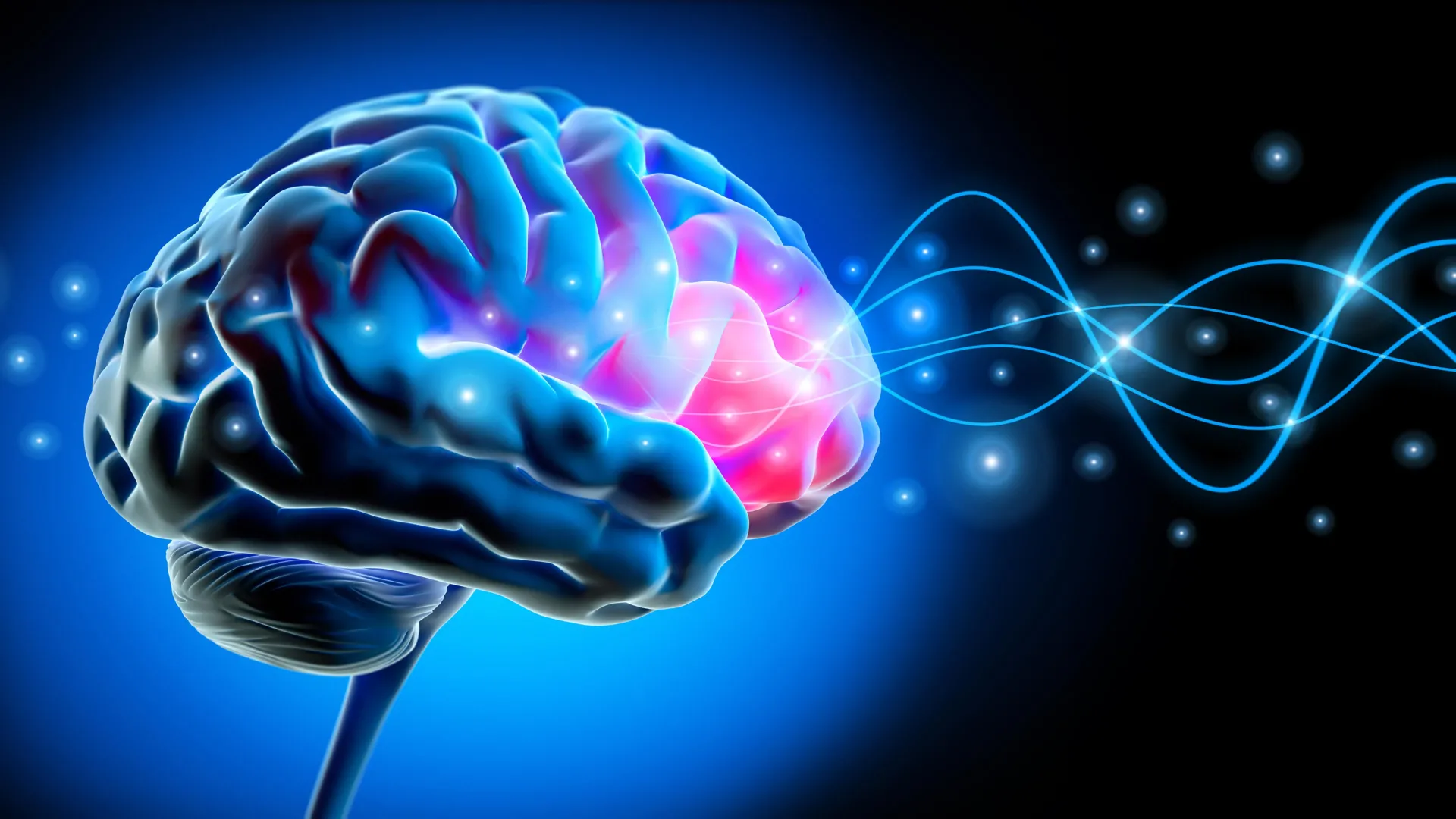
During deep NREM sleep, the human brain undergoes a massive, highly coordinated electrical reset. Vast populations of neurons synchronously transition from an active state to a brief period of inactivity, before firing back into coordinated action. This synchronized physiological shutdown and reactivation generates massive, slow electrical waves. These traveling slow waves typically originate in the frontal cortex—the brain region responsible for higher-order executive functions, decision-making, and memory management—and propagate outward across broad neural networks.
"Each slow wave reflects an enormous population of neurons switching off and back on together," explained Omer Sharon during an overview of the mechanics behind the study. "Our study shows that for memory, it’s critical that these events cascade in sequence across large parts of the brain. This coordinated shutdown has been observed only during deep sleep, and may be one reason why we sleep."
These traveling waves act as a neural transit system, transferring newly acquired, fragile memories from temporary storage in the hippocampus—the brain’s processing hub—to the neocortex for long-term archiving. When this electrical cascade functions smoothly, individuals wake up with sharpened memory retention and enhanced cognitive clarity. When it breaks down, the brain’s filing system falters.
Investigating the Aging Brain: Methodology and Findings
To examine how this delicate neural choreography changes over time, the UC Berkeley research team deployed a multi-layered investigative methodology. The study compared two distinct cohorts: cognitively healthy young adults in their early 20s and a group of older participants ranging in age from their mid-60s to mid-70s.
The researchers utilized high-density electroencephalograms (EEGs) to monitor and record the electrical activity of the participants’ brains while they slept. The physiological contrast between the two age groups was immediate and striking. In the younger cohort, slow waves moved harmoniously in synchronized groups across broad neural landscapes, covering distances roughly equivalent to the width of an adult hand on the scalp. Conversely, in the older participants, these waves typically traveled significantly shorter distances and appeared sporadically and independently.
The research team termed these fragmented, isolated patterns "lonely waves."
To determine whether these altered sleep signatures carried functional consequences for memory retention, the participants were subjected to a rigorous cognitive assessment. In the evening, subjects were tasked with learning pairs of associated words. The following morning, they were tested on their ability to recall those pairs, providing the researchers with a quantifiable metric of overnight memory consolidation.
The empirical data revealed a direct statistical correlation: older participants whose brains produced more isolated, shorter-traveling slow waves consistently demonstrated poorer overnight memory retention.
The Tau Factor: Unveiling Subclinical Pathology
The presence of "lonely waves" and diminished memory performance raised a critical question: what physiological mechanism was driving the breakdown of these synchronized electrical storms?
To answer this, the Berkeley team collaborated with Dr. William Jagust, a professor emeritus of public health and neuroscience at UC Berkeley, utilizing positron emission tomography (PET) scans. By introducing safe radioactive tracers into the bloodstream, the PET imaging illuminated the precise biological activity and distribution of tau proteins within the brains of the older participants.
Tau is a structural protein that, when healthy, stabilizes the internal skeleton of nerve cells. However, in neurodegenerative conditions like Alzheimer’s disease, tau proteins detach, misfold, and clump together into neurofibrillary tangles, progressively choking neurons to death.
The PET imaging revealed a profound association: greater accumulation of tau protein specifically within the frontal cortex was directly linked to the degradation of long-distance traveling slow waves during deep sleep.
Crucially, the study’s subjects were clinically healthy. "These people did not have Alzheimer’s," Sharon emphasized. "They had tau in their brains, but with subclinical impacts. Their memory decline was within the normal range for their age."
This distinction is vital. The findings indicate that Alzheimer’s-related pathology begins accumulating years—potentially decades—before clinical symptoms manifest. Even when memory loss remains within the bounds of what is considered "normal aging," the underlying burden of tau is actively degrading the neural hardware required for optimal sleep-dependent memory processing.
Corroborating Evidence from Independent Cohorts
To ensure the scientific validity and robustness of these findings, the research team sought to replicate their observations using an entirely independent methodology and cohort. Because PET imaging is resource-intensive and restricted to specialized facilities, the Berkeley researchers partnered with neurologist Dr. Yo-El Ju at Washington University in St. Louis.
This collaboration analyzed cerebrospinal fluid (CSF) samples obtained from a separate group of older adults. While spinal fluid analysis cannot pinpoint regional tau accumulation within the frontal cortex in the same manner as a PET scan, it provides an accurate biomarker profile of the central nervous system’s chemical environment.
The results mirrored the imaging data with striking consistency. Older adults whose sleeping brains produced a higher frequency of isolated slow waves exhibited elevated ratios of tau to amyloid-beta—another hallmark protein associated with Alzheimer’s disease—in their spinal fluid.
"It’s collected in a totally different place and in a different cohort, so I think that adds to the strength of the evidence," Sharon noted, highlighting how the convergence of EEG sleep recordings, PET imaging, CSF biomarkers, and behavioral memory tests solidified the study’s conclusions.
Broader Implications and Future Horizons
The implications of this research extend far beyond academic neuroscience, offering a potential framework for future clinical interventions and diagnostic tools.
Historically, cognitive decline in older adults has been viewed as a monolithic, unavoidable consequence of cellular aging. This study demonstrates that individual variability in cognitive aging is heavily influenced by specific neurodegenerative pathologies—namely, localized tau accumulation interrupting global sleep architectures.
"It’s not just about age," Sharon stated. "It’s how much pathology you have in frontal areas where global waves originate."
By establishing that deep sleep disruption serves as a measurable downstream effect of early tau pathology, the research opens exciting new avenues for early detection. Monitoring sleep architecture using non-invasive EEG technology could eventually serve as an accessible, cost-effective biomarker screening tool, alerting physicians to subclinical neurodegenerative changes long before clinical memory deficits emerge.
Furthermore, the findings provide a clear target for future therapeutic development. Interventions aimed at preserving or artificially enhancing slow-wave synchronization during deep sleep could potentially mitigate memory loss in aging populations, slowing the cognitive march of neurodegeneration.
However, significant scientific hurdles remain. While the study definitively establishes a longitudinal correlation between rising tau levels, increasingly lonely sleep waves, and declining episodic memory, causality has not yet been definitively proven.
"We can see tau and lonely waves rising together over time," Sharon cautioned. "What we cannot yet say is which leads—and that question will shape how we design interventions."
Future research must determine whether accumulating tau directly dismantles synchronized sleep waves, whether chronic disruptions in slow-wave sleep accelerate tau pathology, or whether a reciprocal feedback loop drives both processes simultaneously.
As the scientific community races to find effective treatments for Alzheimer’s disease and age-related cognitive decline, the quiet hours of the night—and the synchronized waves pulsing through the sleeping brain—may hold the roadmap to preserving human memory.