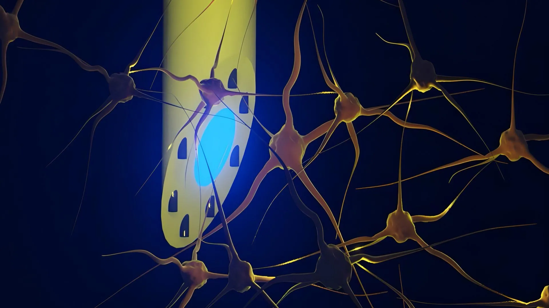
A groundbreaking advancement in neuroengineering has yielded a novel brain implant capable of providing scientists with an exceptionally precise mechanism for studying complex neural pathways and potentially paving the way for advanced treatments for neurological disorders such as epilepsy. Developed by an international consortium of researchers originating from the Technical University of Denmark (DTU), the University of Copenhagen, and University College London, alongside other collaborating academic institutions, the device represents a significant leap forward from conventional rigid silicon-based electrodes and flat-ended optical fibers.
Described in a study recently published in the peer-reviewed scientific journal Advanced Science, the device is formally known as the microfluidic Axialtrode, or mAxialtrode. Measuring less than half a millimeter across and engineered from soft, flexible polymer materials, the needle-thin implant combines multiple functional capabilities into a single, cohesive fiber. This design allows researchers to simultaneously record electrical activity, deliver precise doses of medication, and administer light stimulation across multiple distinct depths and layers of the brain. While the technology is currently positioned as a premier research instrument for investigating neurological phenomena, its developers project that it could eventually transform clinical therapeutic interventions for conditions marked by aberrant electrical activity, such as drug-resistant epilepsy.
The Chronology of the Innovation
The development of the mAxialtrode follows years of incremental progress in the fields of optogenetics, flexible electronics, and microfluidics. Conventional brain-computer interfaces and neural recording tools have historically relied on rigid materials like silicon or heavy metals. While these legacy devices have yielded immense insights into neuroscience over the past several decades, their mechanical rigidity presents a fundamental mismatch with the soft, gel-like consistency of living brain tissue. Over time, the micro-motions of the brain against rigid implants cause chronic inflammation, glial scarring, and the degradation of recorded signals, severely limiting the long-term viability of chronic neural interfaces.
Recognizing these limitations, post-doctoral researcher Kunyang Sui and Associate Professor Christos Markos at DTU conceptualized a softer, multifunctional alternative. By leveraging advanced thermal drawing techniques—a manufacturing process where a macroscopic polymer preform is heated and pulled into an exceptionally fine, microscopic strand with high geometric precision—the team succeeded in producing a flexible fiber that mimics the mechanical compliance of neural tissue.
Following the initial fabrication and structural optimization phases, the engineering team partnered with neurophysiologists, including Associate Professor Rune W. Berg of the University of Copenhagen and Associate Professor Rob C. Wykes of University College London. This multidisciplinary collaboration enabled rigorous in vivo validation. The researchers successfully tested the mAxialtrode in living murine models, connecting the implanted fibers to external optical setups, electrophysiological recording equipment, and miniature fluidic pumps. The successful demonstration of simultaneous multi-site recording, targeted drug delivery, and multi-wavelength optical stimulation in freely moving subjects marked a critical milestone in the project’s timeline, culminating in the recent publication of their findings and the initiation of patent applications for the core technology.
Anatomy of the mAxialtrode
The mAxialtrode distinguishes itself through a sophisticated internal architecture packed into a fraction of a millimeter. At the center of the polymer fiber lies a light-conducting core designed to transmit photons deep into subcortical structures. Encircling this optical core are eight microscopic channels. These microfluidic conduits serve a dual purpose: they are capable of transporting minute volumes of liquid pharmaceuticals or chemical agents, and they can house ultra-thin metal wires dedicated to capturing local field potentials and single-unit electrical activity from surrounding neurons.
The manufacturing technique ensures that the final outer diameter of the fiber remains under 0.5 millimeters. Furthermore, the device features a specially angled distal tip. This geometric optimization reduces the physical footprint of the implant upon insertion, minimizing acute trauma to the cerebral vasculature and parenchyma. Because the polymer materials used are significantly more flexible than traditional glass or silicon probes, the mAxialtrode flexes in tandem with natural brain pulsations. This mechanical compliance drastically reduces the foreign body response, mitigating the inflammatory cascades that typically compromise signal quality over extended recording periods.
Overcoming the Limits of Conventional Optical Fibers
To appreciate the functional leap represented by the mAxialtrode, it is necessary to examine the constraints of current neuroscientific hardware. For years, optogenetics has relied on flat-ended optical fibers fabricated from glass or rigid plastics. These tools have allowed researchers to illuminate specific populations of genetically modified neurons with high temporal resolution. However, traditional fibers suffer from a critical architectural limitation: light emission and tissue interaction occur strictly at the distal terminus, or the "nose" of the fiber.
Consequently, conventional fibers act as single-point sensors and stimulators. To study how information propagates vertically through the layered architecture of the neocortex or between the cortex and deeper structures like the hippocampus, researchers have historically been forced to insert multiple discrete fibers or electrode arrays. This multi-probe approach increases tissue damage, complicates surgical procedures, and introduces spatial registration errors when correlating data across different brain regions.
The mAxialtrode bypasses this bottleneck by providing multiple functional nodes along its longitudinal axis. Because the microfluidic channels and embedded electrical leads can interact with tissue at staggered depths—spanning distances of nearly three millimeters in experimental setups—a single inserted fiber can monitor and modulate distinct neural layers simultaneously. This capability mirrors the distributed, network-level complexity of natural brain function, where cognitive processes, memory consolidation, and pathological states like epileptic seizures depend on rapid, multi-regional communication.
Experimental Validation and In Vivo Performance
During the recent validation phase, the research team implanted the mAxialtrode into the brains of living mice to evaluate its performance under physiological conditions. The subjects were fitted with lightweight assemblies that integrated the flexible fiber with miniature laser diodes, data acquisition systems, and micro-pumps, allowing the animals to move about their enclosures without behavioral impedance or signs of physical distress.
The experimental results confirmed that the implant could reliably deliver localized light stimuli utilizing both blue and red wavelengths, activating targeted optogenetic pathways. Simultaneously, the embedded electrical recording channels captured clear neural signals from both shallow cortical layers and deeper subcortical structures, including the hippocampus. Furthermore, the microfluidic system successfully injected distinct chemical agents at separate depths along the single implant tract, proving that chemical, electrical, and optical modalities can be operated independently or in concert without cross-interference.
Broader Implications and Future Clinical Horizons
While the successful in vivo tests mark a major technical achievement, the research team emphasizes that the mAxialtrode remains firmly within the domain of foundational research. Dr. Kunyang Sui and his colleagues note that translating the technology from laboratory animal models to human clinical applications will require extensive biocompatibility profiling, long-term stability testing, and rigorous evaluation by regulatory bodies such as the U.S. Food and Drug Administration (FDA) or European equivalents.
Nevertheless, the long-term therapeutic implications are profound. Neurological disorders such as epilepsy are frequently characterized by focal disruptions in electrical signaling that cascade into widespread network dysfunctions. Current neurostimulation therapies, such as responsive neurostimulation (RNS) or deep brain stimulation (DBS), typically deliver electrical currents to fixed locations to disrupt seizure activity.
A multifunctional implant like the mAxialtrode opens the door to closed-loop combinatorial therapies. In a hypothetical future clinical scenario, a similar device could continuously monitor neural activity across multiple brain layers to detect the earliest electrophysiological signatures of an oncoming seizure. Upon detection, the implant could autonomously deliver an anticonvulsant medication directly to the precise epileptic focus via its microfluidic channels, while simultaneously applying targeted electrical or optogenetic stimulation to arrest the abnormal neural firing before a clinical seizure manifests. By combining localized chemical delivery with electrical and optical neuromodulation through a single, tissue-friendly implant, medical science may eventually achieve a level of therapeutic precision that minimizes systemic side effects and maximizes clinical efficacy.
As the DTU-led consortium moves forward with patenting the underlying technology and plotting the path toward clinical translation, the mAxialtrode stands as a testament to the convergence of materials science, microfluidics, and neuroengineering—offering a clearer window into the workings of the human brain and a beacon of hope for future neurological interventions.